

The most advanced CBCT technology to acquire volumetric images with a single scan and less radiation dose.
X-VIEW 3D PAN
The X-VIEW 3D system combines the latest advances in digital radiology with high-res 50 Voxel image clarity, an IGZO sensor, and a clean and compact design to obtain high resolution 3D images of the dental structures, soft tissues, nerve paths and bone in the craniofacial region.
FEATURES
Multi-FOV units include FOV's from 5x5cm to 11x14cm


PRECISE DIAGNOSTICS
Images obtained with X-VIEW 3D cone beam CT allow for more precise diagnostics.
LOW RADIATION DOSE
The high frequency generator and the pulsed emission adjust the exposure adapting the dose to the dimensions of the examined area without compromising image quality.
ACCURATE IMAGES
Thanks to its evolved functions, trajectories, and collimations applicable to each exam, images are full of details and contrast
HIGH CONNECTIVITY
The software offers a wide range of tools to manage the images allowing to efficiently save, export and share files.
GO-GREEN AND SAFE PACKAGING
The new detachable column allows the unit to be safely packed in a single triple-wall corrugated box (120 × 80 × 120 cm), using sustainable materials and methods that reduce energy use and environmental impact.
Achieve flawless images with the innovative features integrated into X-VIEW 3D PAN, for outstanding results.
-
3D CBCT RECONSTRUCTION
Even in its Basic Version, without activating additional advanced features, the algorithm surpasses standard reconstruction techniques like filtered back projection (FBP) or Feldkamp (FDK). -
METAL ARTIFACT REDUCTION (MAR)
Activate this option when high-density objects, like metallic components, are present within the scanned field. These objects typically lead to pronounced streaks and dark shadows. -
PATIENT MOTION CORRECTION
Patient movement can lead to the appearance of double edges and blurred structures. This feature identifies and mitigates patient motion, ensuring sharp details and accurate reconstruction of anatomy. -
INCREASED HOMOGENEITY
A dedicated reconstruction kernel tailored for dental scans enhances contrast, resulting in improved homogeneity and reduced brightening at the image periphery. The homogeneity factor determines the distribution of gray and/or color values within the image. Homogeneity is achieved when each pixel in the image shares the same color, while strong contrasts indicate inhomogeneity. -
ADVANCED CALIBRATION
By using a specific geometric calibration tool and software module, it is possible to automatically and accurately calculate all of the scanner’s geometric parameters and compensate for any repeatable mechanical inaccuracies.
-
3D VOLUMETRIC IMAGES WITH VARIABLE FOV
FOV STANDARD DOSE


X-VIEW 3D PAN offers multiple Field of View (FOV) options, while maintaining a standard dose for patient safety.
Experience precise imaging with customizable FOV settings that provide detailed visuals without compromising on radiation exposure.FOV REDUCED DOSE


X-VIEW 3D PAN offers a larger field of view (FOV) for the same radiation dose, effectively reducing the dose per scan.
Benefit from wider image coverage without increasing radiation exposure, ensuring greater diagnostic accuracy with optimal patient safety.X-VIEW 3D PAN allows image acquisition in three different resolution settings:
HD
Ideal for those seeking the highest quality in terms of high definition, although it requires more storage space.
STD
It represents an optimal compromise between image quality and file size.
WLF
A particularly lightweight version offering good image quality.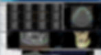

The PC for X-VIEW 3D comes with Deep-View software + Xelis Basic software installed. The computer is configured from the factory in every single detail. The calibration files are already provided, so the user does not have to manually install them or even manipulate the computer with instructions for operation, avoiding complicated calibrations on site, after the installation, or during the unit useful life.
QUICK AND EASY PATIENT POSITIONING


The ergonomic design ensures that the patient is not enforced to use awkward postures and can easily achieve the correct positioning.
The specially designed tools assist patients, during the acquisition process, to maintain their position avoiding errors in the procedures.
X-VIEW 3D Pan adapts to all sizes and types of patients, the linear and open design facilitates access for wheelchair users.
7-INCH COLOUR TOUCHSCREEN


The LCD display enables users to directly interact with the unit.
Menus are very easy to navigate.
Universally recognized icons indicate available functions, parameters, and options.
All instructions are multilingual in a friendly language that guides the user fluently through the different options.
FUNCTIONS


2D PAN FUNCTION
The standard panoramic program provides a precise definition of the dental anatomy in just 15 seconds allowing:
-
Overall evaluation of dentition
-
Examine for intraosseous pathology, such as cysts, tumors, or infections
-
Gross evaluation of temporomandibular joints
-
Evaluation of position of impacted teeth
-
Evaluation of eruption of permanent dentition
-
Dentomaxillofacial trauma
-
Development of disturbances on maxillofacial skeleton


CEPH FUNCTION
24×30 cm AP, LL cephalograms
Carpus images
-
Describes the patient’s dento-facial morphology
-
Quantitatively describes the morphological deviations
-
Obtains orthodontics diagnosis and treatment planning
-
Supports the planning of maxillofacial surgery
-
Elaborates Airway analysis
-
Analyzes the skeletal and dental abnormalities
-
Evaluates treatment results
-
Predicts growth related changes
GO TO THE NEXT LEVEL With Deep-AI

Deep-AI
Deep-AI is not a filter. It is a revolution in panoramic imaging. Sharper images. Smarter diagnostics. Zero effort. Let Deep-AI do the work for you. Deep-AI is the premium module for Deep-View that transforms every 2D panoramic scan into a clearer, more accurate, and diagnosis-ready image.
Unlike traditional filters, Deep-AI automatically analyzes anatomical structures and dental contours. It adjusts brightness, contrast, sharpness, and exposure in real time-fully automated, with no workflow disruption or post-processing

