
.png)
X-VIEW 2D PAN
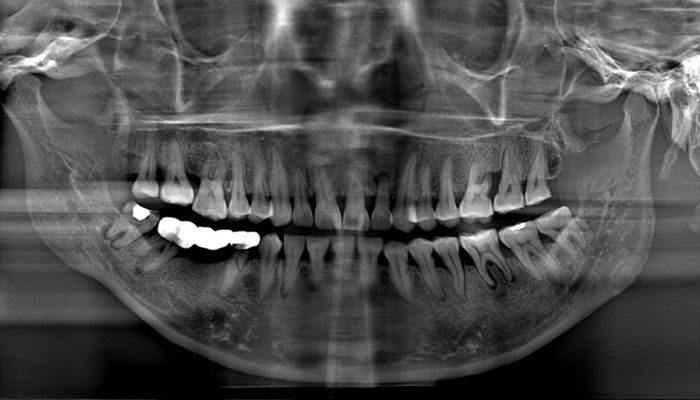
Thanks to the introduction of the most recent technology and the newest features in digital radiology, X-VIEW 2D PAN requires a minimum exposure time to capture clear and contrasted images.
Panoramic images acquired with X-VIEW 2D PAN have a precise focus throughout the entire arch and facilitate the general evaluation of the quantity and quality of bone, dentition and TMJ.
OUR SOFTWARE
Deep-View is a modern imaging software designed by Trident to efficiently acquire, organize, store and share digital images.
The suite is a complete and integrated software for intuitive navigation with several advanced features to obtain and manage thousands of high-definition images for more accurate diagnosis.

EFFICIENT
Save time, optimize files’ management, and speed up the workflow.
EASY TO USE
Run the amazing features of Deep-View in a few simple steps.
MULTIPLATFORM
The interface innovative design makes Deep-View easy to use in multiple devices.
SECURE
Advanced cryptography techniques ensure data protection.
QUICK AND EASY PATIENT POSITIONING


The ergonomic design ensures that the patient is not enforced to use awkward postures and can easily achieve the correct positioning.
The specially designed tools assist patients, during the acquisition process, to maintain their position avoiding errors in the procedures.
X-VIEW 2D adapts to all sizes and types of patients, the linear and open design facilitates access for wheelchair users.
7-INCH COLOUR TOUCHSCREEN


The LCD display enables users to directly interact with the unit.
Menus are very easy to navigate.
Universally recognized icons indicate available functions, parameters, and options.
All instructions are multilingual in a friendly language that guides the user fluently through the different options.
FUNCTIONS


2D PAN FUNCTION
The standard panoramic program provides a precise definition of the dental anatomy in just 15 seconds allowing:
-
Overall evaluation of dentition
-
Examine for intraosseous pathology, such as cysts, tumors, or infections
-
Gross evaluation of temporomandibular joints
-
Evaluation of position of impacted teeth
-
Evaluation of eruption of permanent dentition
-
Dentomaxillofacial trauma
-
Development of disturbances on maxillofacial skeleton


-
Describes the patient’s dento-facial morphology
-
Quantitatively describes the morphological deviations
-
Obtains orthodontics diagnosis and treatment planning
-
Supports the planning of maxillofacial surgery
-
Elaborates Airway analysis
-
Analyzes the skeletal and dental abnormalities
-
Evaluates treatment results
-
Predicts growth related changes
GO TO THE NEXT LEVEL WITH FOCUS

Enhance your professional practice with cutting-edge technology in digital 2D image acquisition. The new X-VIEW 2D PAN FOCUS, equipped with advanced Image Reconstruction Technology, surpasses traditional methods for capturing panoramic images.
FOCUS technology generates high-definition 2D images derived from CBCT technology, applying tomographic techniques to the OPG field. These newly acquired images are exceptionally sharp and clear, eliminating the need for post-processing.
During acquisition, the autofocus function automatically identifies the optimal focus zones, ensuring the best possible sharpness and resolution—delivered quickly and with lower radiation exposure.
The X-VIEW 2D PAN FOCUS meets the highest expectations of healthcare professionals. Its dynamic, user-friendly design and flexible technology consistently deliver excellent results, even in the most complex cases.
No autofocus
Autofocus
